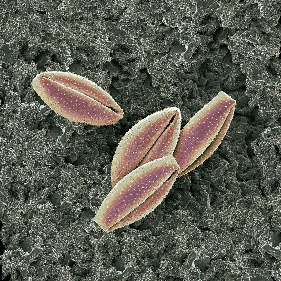
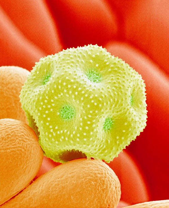
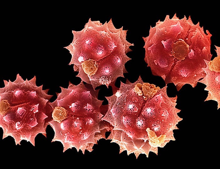
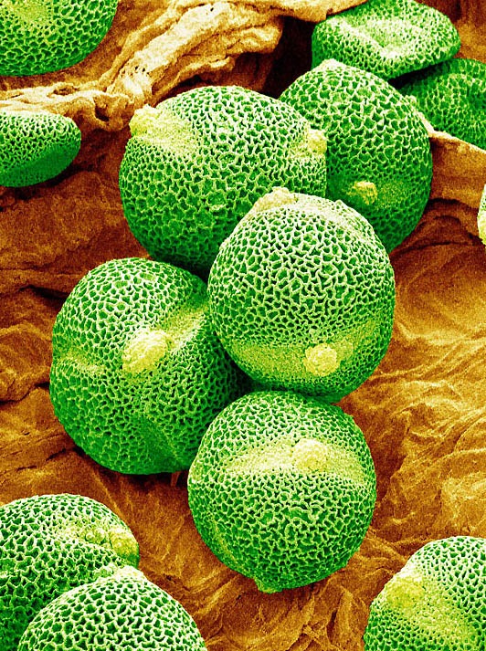
Lily Pollen Grains Photograph by Martin Oeggerli/science Photo Library
Pollen grain recognition is commonly performed by visual inspection by a trained person. An alternative method for visual inspection is automated pollen analysis based on the image analysis technique. Image analysis transfers visual information to mathematical descriptions.
Pollen Grains Under Microscope Amusing
Pollen Grains Under the Microscope - MVA Scientific Consultants Pollen Under the Microscope by Pollen is a fine powdery substance, consisting of microscopic grains released from the male part of a flower or from a male cone. The pollen is transported by the wind, insects, or other animals.
Pollen Grains Under Microscope Amusing
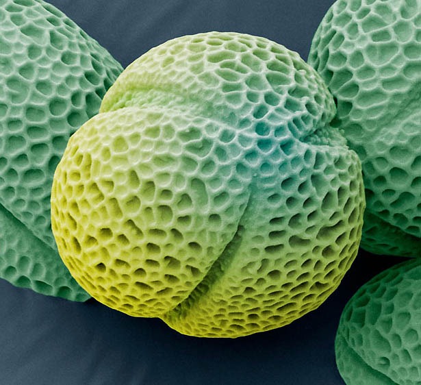
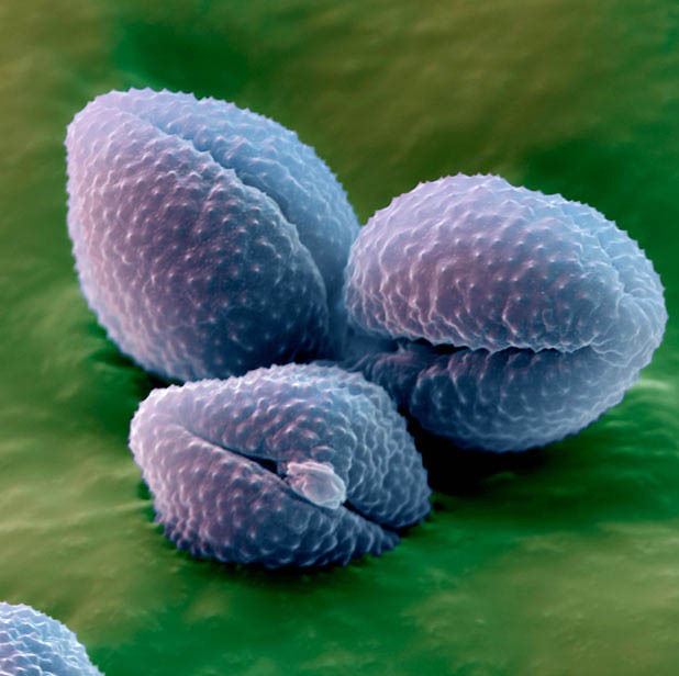
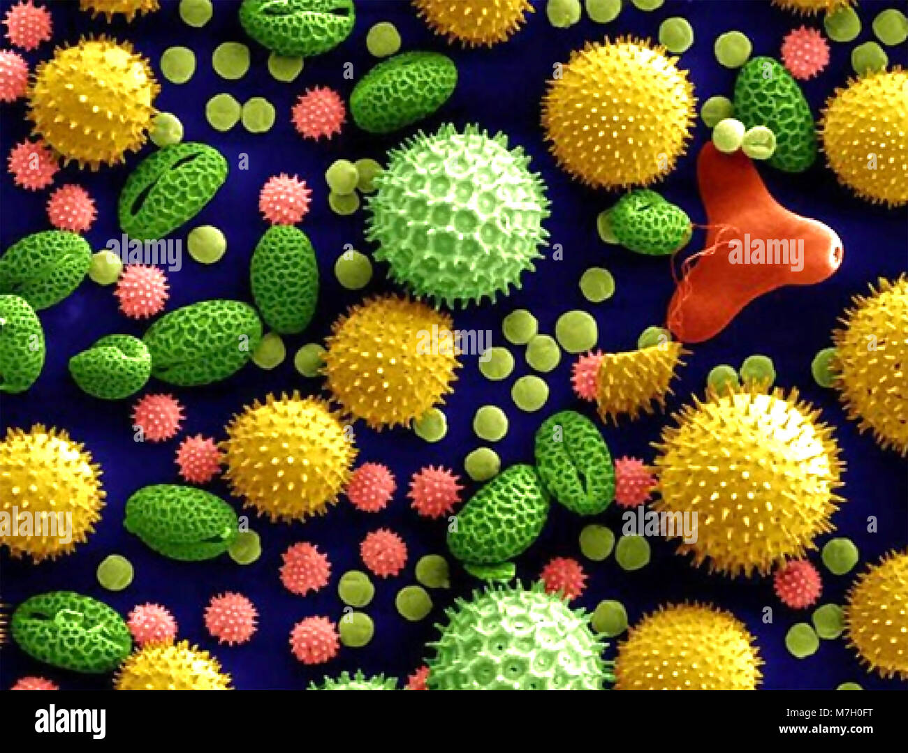
A ctual pollen is merely the best-known subject of study, and the most spectacular. Under a microscope, the individual grains of pollen from different species can look like soccer balls, sponges, padded cushions, coffee beans, or burr balls from the sweetgum tree.
Lily Anther Pollen Microscope Images at Orion Telescopes
This method requires the subsequent counting of the pollen grains under optical microscopy, the most common and economic system to analyze samples, according to a protocol.. Obviously, when moving the position of the sample in the microscope, the pollen grain would appear complete in a new sample in the X or Y direction, and then, it would.
Pollen Grains Under Microscope Amusing
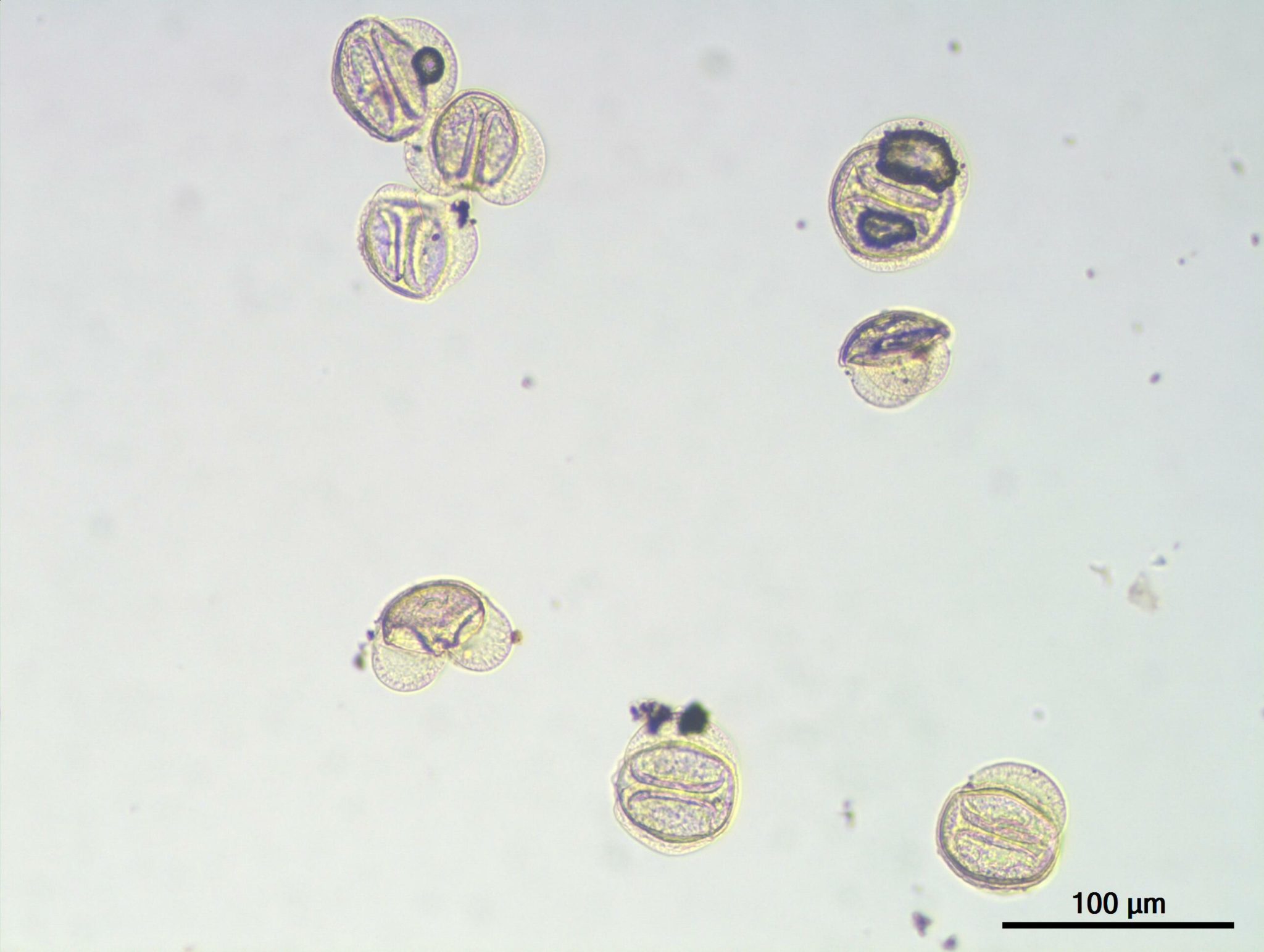
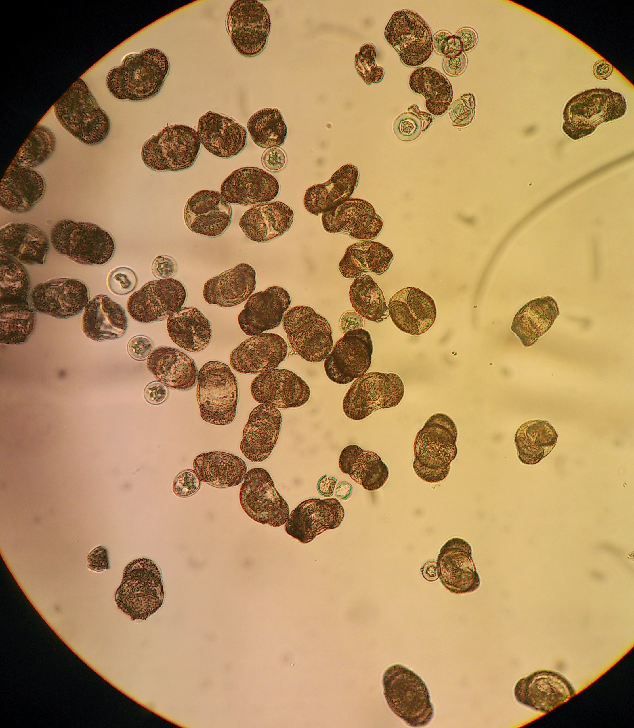
Monocolpate Monocolpate: Pollens with one furrow and without an associated pore or transverse furrow. Monocolpate pollens are found in the Liliaceae (Lily Family) primarily. ( Click here for more images in this category.)
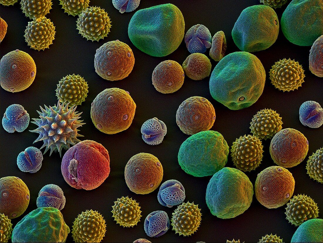
Assortment of pollen grains Bild kaufen 11539239 Science Photo Library
Pollen grain recognition is commonly performed by visual inspection by a trained person. An alternative method for visual inspection is automated pollen analysis based on the image analysis technique. Image analysis transfers visual information to mathematical descriptions.
POLLEN grains under an electron microscope. Photo Courtesy of Dartmouth Electron Microscope
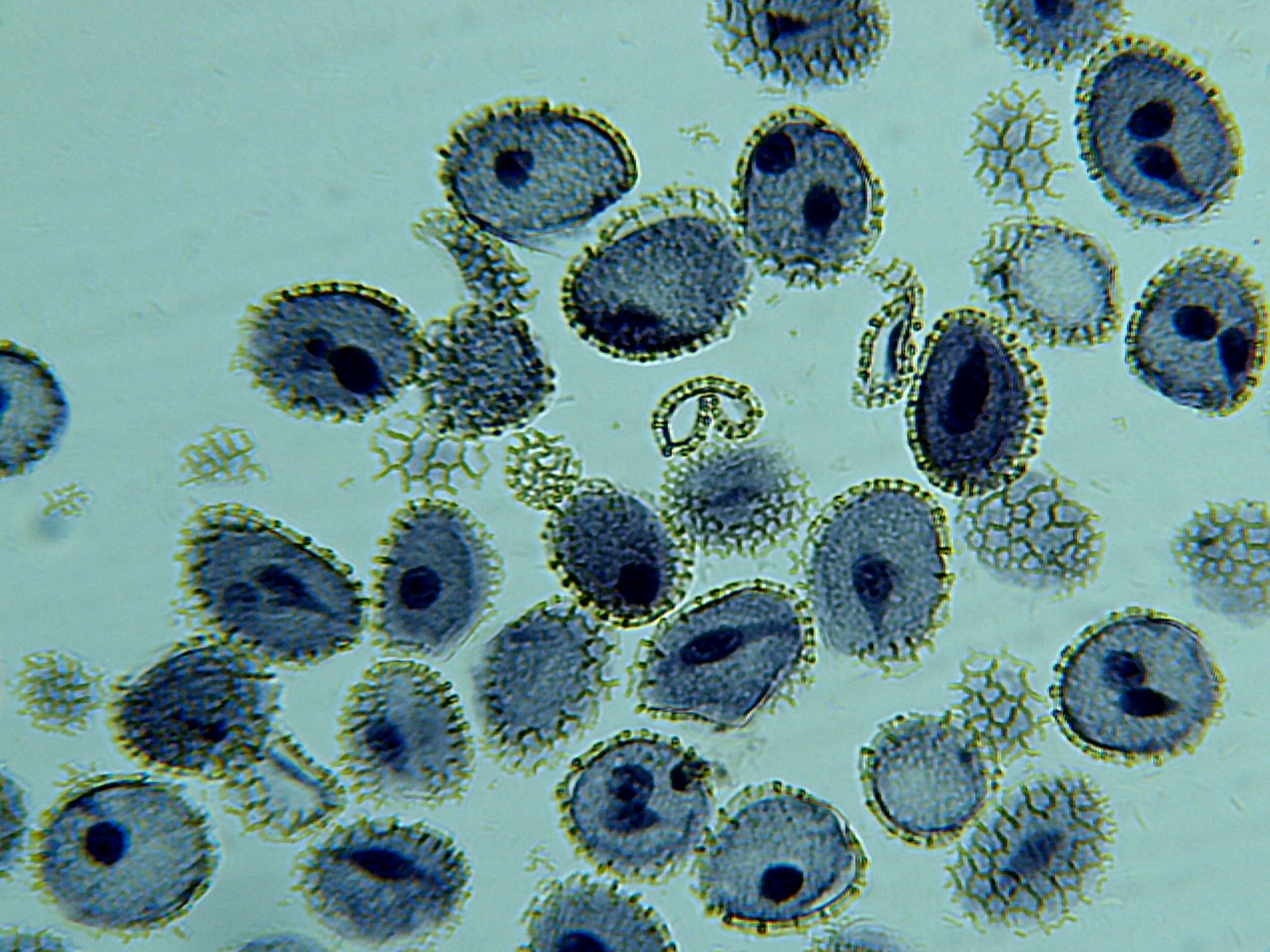
To observe pollen grains under a microscope, follow these steps: 1. Collect the pollen: Choose a flower that is in full bloom and gently tap the anthers to release the pollen grains onto a clean glass slide. Alternatively, you can use a fine brush or forceps to collect the pollen from the anthers. 2.

Microscope World Blog Pollen under the Microscope
We also checked non-acetolyzsed pollen grains from both cultivars under a light microscope, to confirm the preparation method had not significantly affected pollen morphology (Supplementary Fig. S6).
Pollen Grains Under the Microscope MVA Scientific Consultants
Pollen Under The Microscope Methods, Techniques and Observations What is Pollen? Pollen is a small grain that consists of a few cells. To the naked eye it appears as a yellowish (pale yellow) dust-like substance that is either dispersed by wind or insects.
Pollen Grains Under Microscope Amusing
1. Introduction The monitoring of pollen and fungal spores concentra-tions allows the detection and the quantification of aller-genic species [18] and of potential infectious diseases both on humans [9] and plants [21].
A group of pollen grains under the microscope pics
Under The Microscope: Pollen Pollen is flower sperm, and climate change is making more of it every season. Not a great situation for allergy sufferers. Ada McVean B.Sc. | 11 Mar 2019 Environment Grains of pollen actually produce the male gametes (sperm cells) of flowering plants.

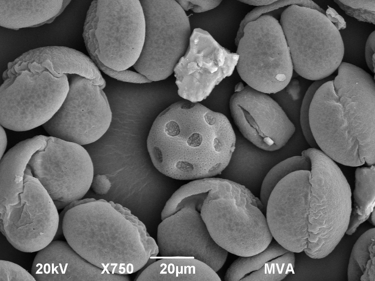
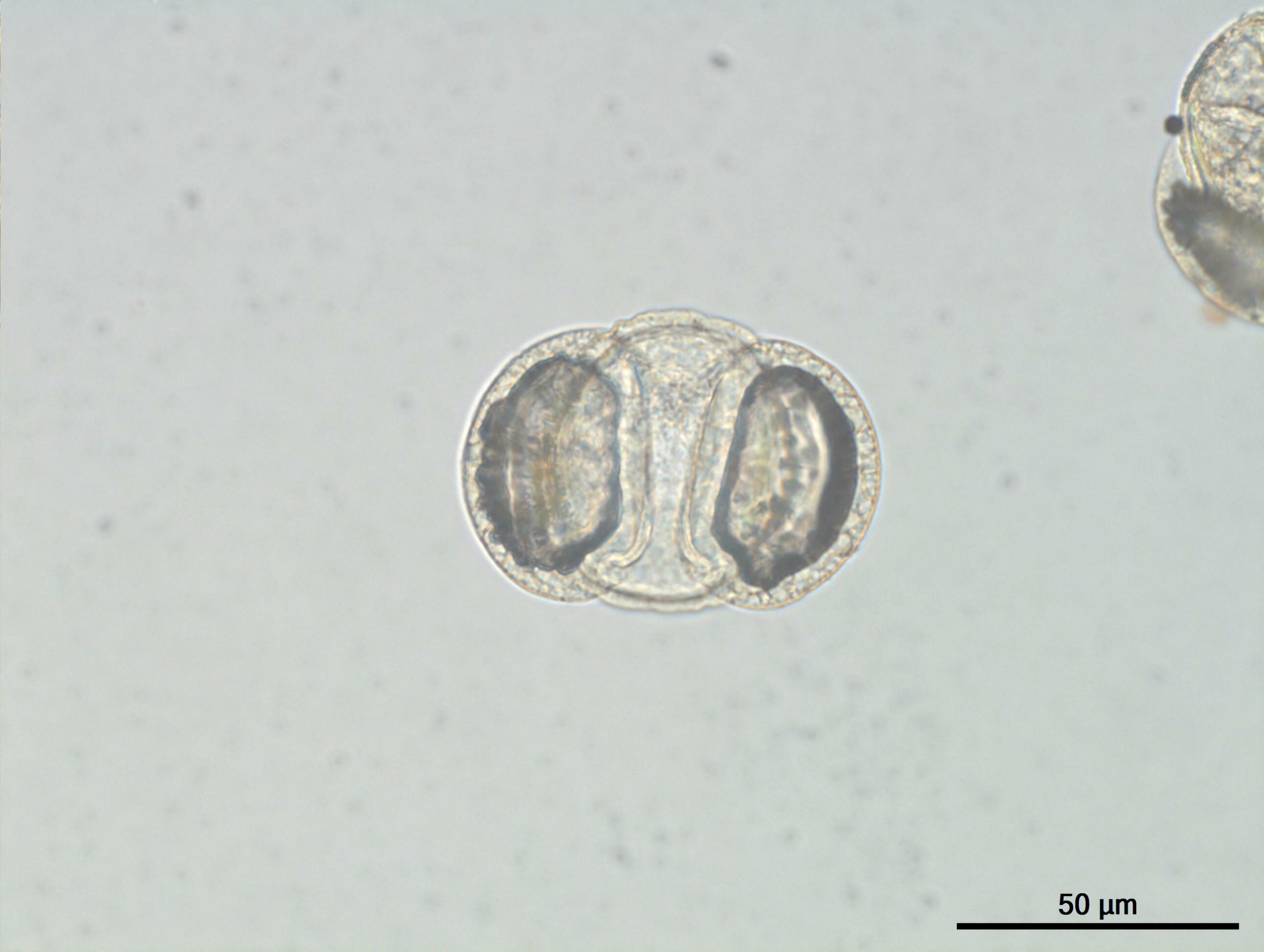
Lily Anther, Mature Pollen Grains, c.s., 12 µm Microscope Slide Carolina Biological Supply
Additionally, given their remarkably symmetrical structure and surface patterns, fresh and preserved pollen grains are readily recognizable under the microscope. Characteristics such as the exine sculpturing and the size and number of apertures through which the pollen tubes grow are useful as taxonomic tools. The structure of a pollen grain is.
PollChem Ecological and Environmental Change Research Group UiB
The largest pollen grains are around 200 µm in diameter, and the smallest are less than 10 µm, so you need a microscope to study them. At 100× you can see the grains, at 400× you can start to see details, and at 1000-1500× you can see enough details to make a good drawing.
Pollen Grains Under the Microscope MVA Scientific Consultants
1. Introduction The analysis of natural microscopic images is the basis of many technological developments in fields such as medicine or biology [ 1, 2, 3 ]. The automatic detection of objects in images captured with an automated microscope is a challenge for many types of natural samples due to the variability of the environment.
Pollen Grains Under Microscope Amusing
Pollen grains are produced by meiosis of microspore mother cells that are located along the inner edge of the anther sacs. [In this figure] The anatomy of a flower. The stamen is a male part of the flower. The stamen contains filament and anther where the pollens are produced. The stigma is a female part of the flower. The structure of pollen
Pollen Grains Under the Microscope MVA Scientific Consultants
Analysis of pollen material obtained from the Hirst-type apparatus, which is a tedious and labor-intensive process, is usually performed by hand under a microscope by specialists in palynology. This research evaluated the automatic analysis of pollen material performed based on digital microscopic photos.